News Revolutionizing Battlefield Medicine: Augmented Reality for Lifesaving Trauma Care

Home / Articles / External Non-Government

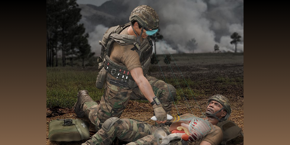
APL researchers are using a statistical shape atlas — a detailed map of variations in human anatomy — to predict the likely location of internal organs based on external body landmarks. This predictive modeling is aided by augmented reality technology, allowing medics to view an overlay of the patient’s internal anatomy directly on their body. Credit: Johns Hopkins APL/Kevin McCafferty
APL researchers are using a statistical shape atlas — a detailed map of variations in human anatomy — to predict the likely location of internal organs based on external body landmarks. This predictive modeling is aided by augmented reality technology, allowing medics to view an overlay of the patient’s internal anatomy directly on their body (credit: Johns Hopkins APL/Kevin McCafferty).

June 4, 2025 | Originally published by Johns Hopkins Applied Physics Laboratory on February 13, 2025

In remote conflict zones and disaster-stricken areas, the nearest hospital is often hundreds of miles away. Medical teams face the tough task of providing critical care — with limited resources and while under constant threat — to casualties with wide-ranging medical needs.

Researchers at the Johns Hopkins Applied Physics Laboratory (APL) in Laurel, Maryland, are leveraging the power of augmented reality (AR), predictive anatomy visualization, and artificial intelligence to provide field medics with a tool to deliver lifesaving care by helping them clearly visualize where internal organs are situated inside their patients’ bodies.

Focus Areas

Want to find out more about this topic?

Request a FREE Technical Inquiry!